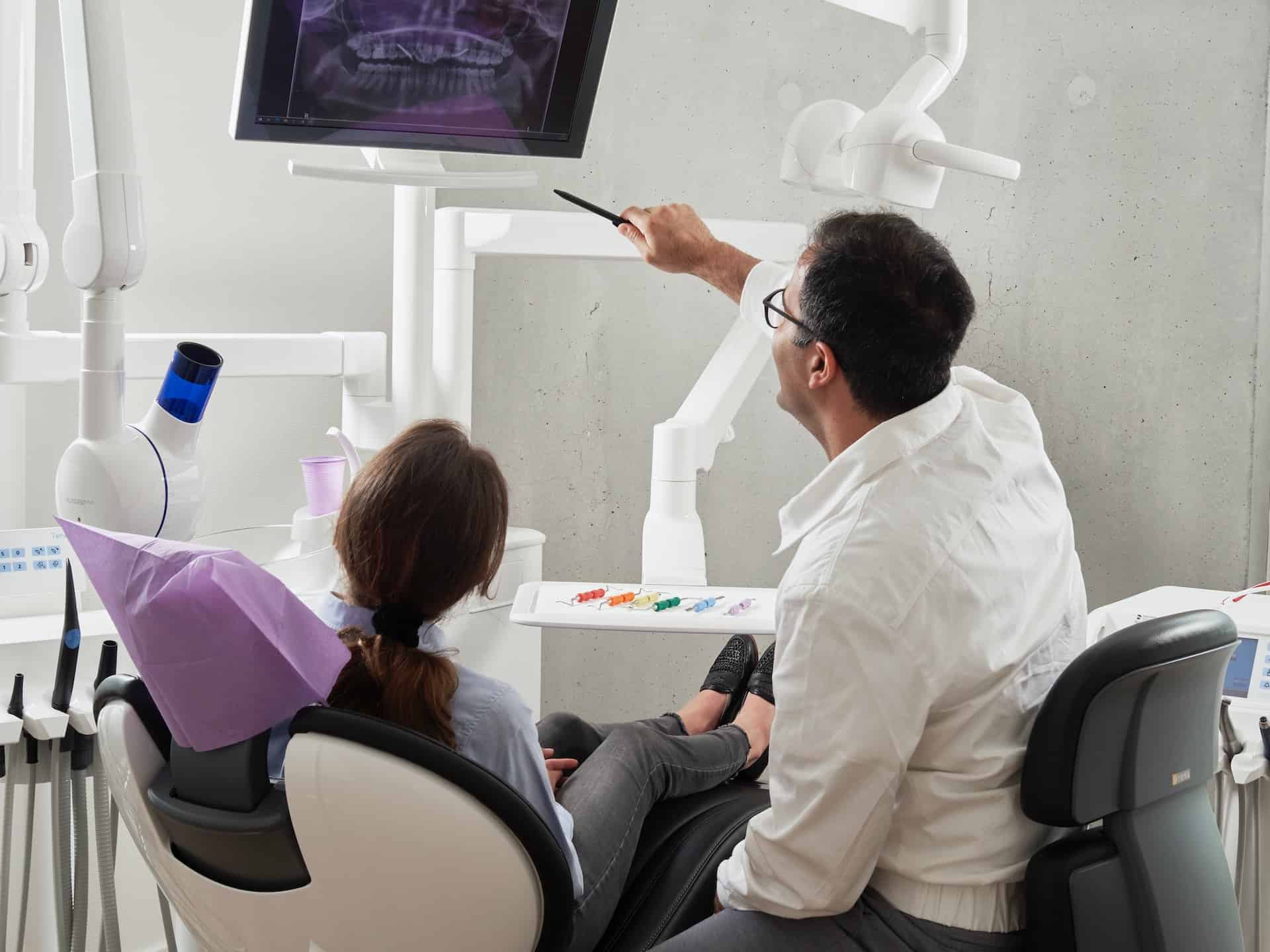
Aligners, dentures, and implants are inseparable parts of dentistry as they offer effective ways to replace missing teeth or support broken ones. Not so long ago, using physical impressions was the primary method for doctors to make a model of the missing teeth, which often was time-consuming, uncomfortable, and somewhat inaccurate. As the demand grows for such tools, many clinics and dental offices turn to dental management software and the technologies it provides. Dental clinics are on the fence about adopting such tools as they do not know how they work and what benefits they offer. The software works by taking multiple X-ray images of the patient’s mouth from different angles, which are then combined to create a 3D model of the teeth, gums, and jawbone. This model can be used to analyze the patient’s dental anatomy, plan treatments, and even simulate the outcome of different procedures. Some benefits of dental 3D imaging software include the following:
– Better diagnosis: The 3D images generated by the software provide a more comprehensive view of the patient’s dental anatomy, allowing for a more accurate diagnosis of dental conditions.
– Improved treatment planning: Dental professionals can use the software to plan and visualize complex procedures, such as dental implants or orthodontic treatments.
– Increased patient education: The 3D models can be shared with patients, allowing them better to understand their dental condition and the recommended treatment options.
– Reduced radiation exposure: While traditional dental X-rays expose patients to small amounts of radiation, 3D imaging software uses lower radiation doses and requires fewer X-rays.
Software for dental offices: ways to improve intraoral scanning and implanting
Dentists have traditionally used alginate materials to create fractured or missing teeth replicas. The procedure can be tedious, time-consuming, and agony for most patients since they must press tightly on the material to obtain accurate impressions. If the impressions are not right, the process has to be repeated.
The scanner spotlights the area while sensors take photos to produce a 3D digital model. Since they are faster, intraoral scanners save time and money when capturing impressions as they reduce chair time. Following are the reasons why dental 3D scanning software is in demand:
– Improved Accuracy: intraoral scans provide highly accurate digital images of the teeth and surrounding tissues, resulting in a more precise fit of the final restoration. Unlike traditional impressions, which can be distorted during pouring, digital scans are not subject to human error.
– Time Savings: intraoral scans can be completed more quickly than traditional impressions, saving patients and dentists time. There is no need to wait for the material to be set, and the process of capturing the images is more efficient.
– Increased Patient Comfort: many patients find the traditional impression process uncomfortable or unpleasant due to the materials used and the amount of time required to complete the impression. Intraoral scans are more comfortable as they involve no physical contact with the patient’s mouth.
– Improved Communication: intraoral scans allow dentists to share detailed 3D images with dental laboratories, reducing the need for physical models to be shipped between locations. This can improve communication, reduce errors, and speed up fabrication.
– Reduced Waste: traditional impressions require using materials such as silicone and polyether, which can generate waste and contribute to environmental concerns. In contrast, digital scans are more environmentally friendly, as they do not require these materials.
Dental implants and prosthodontics software
Accuracy is vital to any implant placement as the doctor cannot initiate the procedure without understanding the jaw area with precise teeth positions, bone density, tissues, and sinus cavities. Here, a prosthodontics solution can gather the data and capture a clear picture of the patient’s jaw by scanning the area. This means that implants are inserted precisely in the preset location. This improves the procedure’s safety, predictability, speed, and comfort while also lowering the necessity for bone transplants. It also helps put implants in places impossible before, making them effective for patients with significant bone loss. In fact, some studies suggested that using an intraoral scanner can make a significant difference in the accuracy of the operation. After gathering all required data, the dental image software creates an accurate 3D model of the jaw, allowing the doctors to identify the ideal position for implant insertion.
Prosthodontics software can improve patient care quality by reducing the risk of error in the design and fabrication process and enabling prosthodontists to achieve more predictable and aesthetic outcomes. It can also streamline workflows, reduce treatment times, and enhance communication between prosthodontists and other dental team members.
The digital workflow for creating dental models: how the modernization will look like?
Dental experts now have access to highly advanced intraoral scanners that can quickly capture digital images of a patient’s jaw with great precision. This technology allows dentists to create a flawless 3D digital imprint of the patient’s teeth and surrounding tissues in under a minute, using a six-step digital workflow:
1. Intraoral Scanning. The process begins with capturing digital impressions of the patient’s teeth and surrounding tissues using an intraoral scanner.
2. Digital Model Creation. The digital impressions are then used to create a 3D digital model of the patient’s teeth and gums. The model can be manipulated on a computer screen to view the teeth from different angles and identify any areas of concern.
3. Virtual Treatment Planning. With the digital model, the dentist can virtually plan the treatment, select the type of restoration, tooth position, and shape, and simulate the final result.
4. Computer-Aided Design (CAD). Next, using specialized software, the dentist designs the final prosthesis, such as a crown or bridge, by digitally sculpting it on the 3D model.
5. Computer-Aided Manufacturing (CAM). The CAD design is sent to a milling machine or 3D printer that creates the final restoration using the chosen material.
6. Finishing and Polishing. The restoration is then finished and polished to ensure it looks and feels natural.
Utilizing digital workflows to create dental models can bring several benefits to the treatment process and help dentists streamline the process, reducing the need for multiple appointments and ultimately saving time for both the dentist and the patient.
Moreover, digital workflows can improve communication and collaboration between dentists, technicians, and patients. The ability to manipulate 3D digital models on a computer screen can help dentists, and patients better visualize treatment options, leading to a more informed decision-making process. Ultimately, this can result in better treatment outcomes and a more positive experience for patients.
